Welcome to DU!
The truly grassroots left-of-center political community where regular people, not algorithms, drive the discussions and set the standards.
Join the community:
Create a free account
Support DU (and get rid of ads!):
Become a Star Member
Latest Breaking News
Editorials & Other Articles
General Discussion
The DU Lounge
All Forums
Issue Forums
Culture Forums
Alliance Forums
Region Forums
Support Forums
Help & Search
The Fly: The first full map of an animal brain has been completed. [View all]
It may sound silly to some, but this is a remarkable scientific achievement, a full map of a fly brain, the "connectome." This comes from the news section at the scientific journal Nature and came in on my Nature Briefing news feed.
Gigantic map of fly brain is a first for a complex animal
Subtitle:
Fruit fly ‘connectome’ will help researchers to study how the brain works, and could further understanding of neurological diseases.
By Miryam Naddaf Nature 10 Mar 2023
Some excerpts:
Scientists have generated the first complete map of the brain of a small insect, including all of its neurons and connecting synapses.
The research, published on 9 March in Science1, provides a brain-wiring diagram known as the connectome of a complex animal for the first time — the fruit fly Drosophila melanogaster. The map shows all 3,016 neurons and 548,000 synapses tightly packed in a young Drosophila’s brain, which is smaller than a poppy seed.
The map is a milestone in understanding how the brain processes the flow of sensory information and translates it into action. “Now we have a reference brain,” says Marta Zlatic, a neuroscientist at the University of Cambridge, UK, and co-author of the paper. “We can look at what happens to connectivity in models of Alzheimer’s and Parkinson’s diseases and of any degenerative disease.”
Ideal model
Until now, scientists had mapped the connectomes of only the worms Caenorhabditis elegans and Platynereis dumerilii, and the larva of the sea squirt Ciona intestinalis. Drosophila was an ideal model for connectome studies, because scientists have already sequenced its genome, and the larvae have transparent bodies. Fruit flies also exhibit sophisticated behaviours — including learning, navigating landscapes, processing smells and weighing the risks and benefits of an action. “Its size is manageable for current technology,” says Chung-Chuang Lo, a computational neuroscientist at the National Tsing Hua University in Hsinchu, Taiwan.
“If you had asked me in the Eighties, when the C. elegans work was being done, about this project in the fruit fly, it would have been impossible,” says Albert Cardona, a neuroscientist at the University of Cambridge and co-author of the paper...
...The wiring diagram showed that the insect’s brain was multilayered, with pathways of varying lengths connecting brain inputs and brain outputs.
It is “a nice, nested structure”, says Michael Winding, a neuroscientist at the University of Cambridge and co-author of the paper. But some of the brain networks have shortcuts, skipping layers. The authors suggest that such shortcuts increase the brain’s computational capacity and compensate for the limited number of neurons.
Your brain expands and shrinks over time — these charts show how
The team also found that 41% of the brain neurons form ‘recurrent loops’, providing feedback to their upstream partners. These shortcuts and loops resemble state-of-the-art artificial neural networks that are being used in artificial-intelligence research. “It’s interesting that the computer-science field is converging onto what evolution has discovered,” says Cardona...
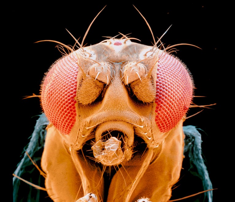
The article is not complete without a picture of the fly:
 ?as=webp
The full scientific article in Science is found here:
The connectome of an insect brain, Michael Winding, Benjamin D. Pedigo, Christopher L. Barnes, Heather G. Patsolic, Youngser Park, Tom Kazimiers, Akira Fushiki, Ingrid V. Andrade, Avinash Khandelwal, Javier Valdes Aleman, Feng Li, Nadine Randel, Elizabeth Barsotti, Ana Correia, Richard D. Fetter, Volker Hartenstein, Carey E. Priebe, Joshua T. Vogelstein, Albert Cardona, Marta Zlatic Science, 379, 6636 (2023) eadd9330
An image from the full paper:

The caption:
Fig. 1. Comprehensive reconstruction of a Drosophila larva brain.
(A) Morphology of differentiated brain neurons in the CNS of a Drosophila larva. (B) Most (>99%) of neurons were reconstructed to completion, defined by reconstruction of all terminal branches (see Methods) and no data quality issues preventing identification of axons and dendrites. Pre- and postsynaptic sites were considered complete when connected to a brain neuron or ascending arbors from neurons outside the brain. (C) Left and right homologous neuron pairs were identified using an automated graph matching with manual proofreading. There was no clear partner for 14 neurons based on this workflow (unpaired), along with 176 unpaired KCs in the learning and memory center. (D and E) Schematic overview of brain structure. Brain inputs include SNs, which directly synapse onto brain neurons, and ANs from VNC segment A1, which receive direct or polysynaptic input from A1 sensories (see fig. S2). Brain interneurons transmit these input signals to output neurons: DNs to the subesophageal zone (SEZ) (DNSEZ), DNs to the VNC (DNVNC), and ring gland neurons (RGN). (F to H) Cell classes in the brain. Some interneurons belong to multiple classes, but are displayed as mutually exclusive for plotting expedience (see fig. S4). Note that some previously reconstructed interneurons (40 total) and output neurons (6 total) are included in the barplots but are not brain neurons per se and not included in counts. There were 20 brain output neurons with known cell classes that were therefore also included in (G).
(A) Morphology of differentiated brain neurons in the CNS of a Drosophila larva. (B) Most (>99%) of neurons were reconstructed to completion, defined by reconstruction of all terminal branches (see Methods) and no data quality issues preventing identification of axons and dendrites. Pre- and postsynaptic sites were considered complete when connected to a brain neuron or ascending arbors from neurons outside the brain. (C) Left and right homologous neuron pairs were identified using an automated graph matching with manual proofreading. There was no clear partner for 14 neurons based on this workflow (unpaired), along with 176 unpaired KCs in the learning and memory center. (D and E) Schematic overview of brain structure. Brain inputs include SNs, which directly synapse onto brain neurons, and ANs from VNC segment A1, which receive direct or polysynaptic input from A1 sensories (see fig. S2). Brain interneurons transmit these input signals to output neurons: DNs to the subesophageal zone (SEZ) (DNSEZ), DNs to the VNC (DNVNC), and ring gland neurons (RGN). (F to H) Cell classes in the brain. Some interneurons belong to multiple classes, but are displayed as mutually exclusive for plotting expedience (see fig. S4). Note that some previously reconstructed interneurons (40 total) and output neurons (6 total) are included in the barplots but are not brain neurons per se and not included in counts. There were 20 brain output neurons with known cell classes that were therefore also included in (G).
Cool.
Have a nice evening.
4 replies
 = new reply since forum marked as read
Highlight:
NoneDon't highlight anything
5 newestHighlight 5 most recent replies
Maybe because they needed something that had more than two working nuerons?
cstanleytech
Mar 2023
#4